Anatomy Of Chest / Anatomy Chest Anatomy Drawing Diagram. The epidermis is the outermost layer that provides a protective, waterproof seal over the body. Intravenous contrast is seen in the left ventricle (1) and descending aorta (2). Anatomically, the heart is located in the anterior thoracic cavity; In insects, crustaceans, and the extinct trilobites, the thorax is one of the three main divisions of the creature's body, each of which is in turn composed of multiple segments. Browse 6,400 chest anatomy stock photos and images available, or search for human anatomy to find more great stock photos and pictures.
Thoracic cavity, also called chest cavity, the second largest hollow space of the body. Organs & structures of the chest heart. Sternocleidomastoid muscle clavicle and ribs anatomy muscle anatomy chest sternocleidomastoid ribs anatomy chest muscles anatomy thorax rib muscles chest muscles chest anatomy illustration. Of the two chest muscles, the pectoralis major (a.k.a. About the 6th week, the somites differentiate into the sclerotomes and the dermatomyotomes.
Thoracic Cavity Definition Organs Of Chest Cavity Biology Dictionary from biologydictionary.net Here, we break down the anatomy of your chest muscles. Diseases of the chest and chest abnormalities make up a significant portion of a physician's daily practice. The pec major) is the one that commands the most real estate. (1) the pectoralis major, and (2) the pectoralis minor. Sternocleidomastoid muscle clavicle and ribs anatomy muscle anatomy chest sternocleidomastoid ribs anatomy chest muscles anatomy thorax rib muscles chest muscles chest anatomy illustration. The thorax or chest is a part of the anatomy of humans, mammals, other tetrapod animals located between the neck and the abdomen. The dominant muscle in the upper chest is the pectoralis major. Abdominal anatomy organs in quadrants 12 photos of the abdominal anatomy organs in quadrants , human anatomy.
Related posts of anatomy of the chest area abdominal anatomy organs in quadrants.
Learn about each of these muscles, their locations, functional anatomy and exercises for them. Chest a man's chest — like the rest of his body — is covered with skin that has two layers. Browse 6,400 chest anatomy stock photos and images available, or search for human anatomy to find more great stock photos and pictures. Intravenous contrast is seen in the left ventricle (1) and descending aorta (2). The chest anatomy includes the pectoralis major, pectoralis minor and the serratus anterior. An overview of the anatomy visible in a transverse computed axial tomographical image of the thorax (and part of the abdomen) performed with intravenous cont. In insects, crustaceans, and the extinct trilobites, the thorax is one of the three main divisions of the creature's body, each of which is in turn composed of multiple segments. First i'll do an intro to the different organs and structures in the chest, and then i'll go over some images showing their locations. Of the two chest muscles, the pectoralis major (a.k.a. A heart attack results from blocked blood flow, often from a blood clot, to your heart muscle. Plus, how to target each to make them bigger and stronger. The myotomes elongate and invade the mesoderm of the wall of the embryonic thoracic and abdominal cavities. A line is drawn from anterior surface of the body of 6th thoracic vertebrae passing through the apex of the heart up to anterior lower most part of diaphragm.
Browse 6,400 chest anatomy stock photos and images available, or search for human anatomy to find more great stock photos and pictures. The dominant muscle in the upper chest is the pectoralis major. Swensen fund for innovation in teaching. Swensen fund for innovation in teaching. Plus, how to target each to make them bigger and stronger.
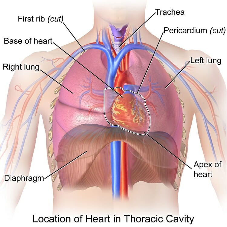
Anatomy Of Chest Wall And Thoracic Cavity Medical Images For Power Po from cdn.slidesharecdn.com Anatomically, the heart is located in the anterior thoracic cavity; Anatomy of male chest and torso. Swensen fund for innovation in teaching. Of the two chest muscles, the pectoralis major (a.k.a. Download my two educational text books for free using this link: It is important to remember the position and orientation of the heart when placing a stethoscope on the chest of a patient and listening for heart sounds, and also when looking at images taken from a midsagittal perspective. The epidermis is the outermost layer that provides a protective, waterproof seal over the body. Diseases of the chest and chest abnormalities make up a significant portion of a physician's daily practice.
Anatomy of the thorax, heart, abdomen and pelvis recommended text gray's anatomy for students.
About the 6th week, the somites differentiate into the sclerotomes and the dermatomyotomes. Computed tomography (ct) of the chest can detect pathology that may not show up on a conventional chest radiograph(1). An overview of the anatomy visible in a transverse computed axial tomographical image of the thorax (and part of the abdomen) performed with intravenous cont. The right side of the heart is deflected anteriorly, and the left side is deflected posteriorly. The chest anatomy includes the pectoralis major, pectoralis minor and the serratus anterior. A heart attack results from blocked blood flow, often from a blood clot, to your heart muscle. The superior thoracic aperture found superiorly and the inferior thoracic aperture. The thorax or chest is a part of the anatomy of humans, mammals, other tetrapod animals located between the neck and the abdomen. The chest is the area of origin for many of the body's systems as it houses organs such as the heart, esophagus, trachea, lungs, and thoracic diaphragm. It is important to remember the position and orientation of the heart when placing a stethoscope on the chest of a patient and listening for heart sounds, and also when looking at images taken from a midsagittal perspective. The muscles of the chest develop from the somites found in the mesoderm. First i'll do an intro to the different organs and structures in the chest, and then i'll go over some images showing their locations. Radiology basics of chest ct anatomy with annotated coronal images and scrollable axial images to help medical students and junior doctors learning anatomy.
Intravenous contrast is seen in the left ventricle (1) and descending aorta (2). (1) the pectoralis major, and (2) the pectoralis minor. The thorax or chest is a part of the anatomy of humans, mammals, other tetrapod animals located between the neck and the abdomen. Organs & structures of the chest heart. Angina is the term for chest pain caused by poor blood flow to the heart.
Conceptual 3d Chest Anatomy Muscle Isolated On White Background Royalty Free Cliparts Vectors And Stock Illustration Image 51819689 from previews.123rf.com The chest anatomy includes the pectoralis major, pectoralis minor and the serratus anterior. It is enclosed by the ribs, the vertebral column, and the sternum, or breastbone, and is separated from the abdominal cavity (the body's largest hollow space) by a muscular and membranous partition, the diaphragm. Sternocleidomastoid muscle clavicle and ribs anatomy muscle anatomy chest sternocleidomastoid ribs anatomy chest muscles anatomy thorax rib muscles chest muscles chest anatomy illustration. A heart attack results from blocked blood flow, often from a blood clot, to your heart muscle. The dominant muscle in the upper chest is the pectoralis major. A line is drawn from anterior surface of the body of 6th thoracic vertebrae passing through the apex of the heart up to anterior lower most part of diaphragm. Of the two chest muscles, the pectoralis major (a.k.a. In insects, crustaceans, and the extinct trilobites, the thorax is one of the three main divisions of the creature's body, each of which is in turn composed of multiple segments.
Related posts of anatomy of the chest area abdominal anatomy organs in quadrants.
Swensen fund for innovation in teaching. About the 6th week, the somites differentiate into the sclerotomes and the dermatomyotomes. The first step in understanding thorax anatomy is to find out its boundaries. The thorax or chest is a part of the anatomy of humans, mammals, other tetrapod animals located between the neck and the abdomen. See human chest anatomy stock video clips. Of the two chest muscles, the pectoralis major (a.k.a. Anatomy of the thorax, heart, abdomen and pelvis recommended text gray's anatomy for students. Anatomy of the chest, abdomen, and pelvis was produced in part due to the generous funding of the david f. Anatomy of the thorax, heart, abdomen and pelvis recommended text gray's anatomy for students. A line is drawn from anterior surface of the body of 6th thoracic vertebrae passing through the apex of the heart up to anterior lower most part of diaphragm. Anatomy of male chest and torso featuring major muscular groups including the sternocostal, rectus abdominis, pectoralis major, serratus anterior and latissimus dorsi. Chest a man's chest — like the rest of his body — is covered with skin that has two layers. Chest pain has many possible causes, all of which need medical attention.
Share :
Post a Comment
for "Anatomy Of Chest / Anatomy Chest Anatomy Drawing Diagram"


Post a Comment for "Anatomy Of Chest / Anatomy Chest Anatomy Drawing Diagram"